facs flow cytometry protocol
Centrifuge fixed cells and resuspend pellet in 1 mL of PBS. Propidium Iodide Cell Cycle Staining.
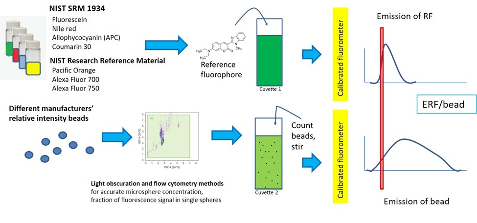
Quantitative Flow Cytometry Measurements Nist
ZERO BIAS - scores article reviews.
. Buy Intracellular Flow Cytometry Reagents conjugated monoclonal antibodies at Santa Cruz. Super Bright Staining Buffer protocol. Ad Request a quote and see how Agilent has advanced the boundaries of flow cytometry.
The following flow cytometry. True-Nuclear Transcription Factor Staining Protocol for 5mL Tubes. Cell Preparation for Flow Cytometry Protocols Invitrogen eBioscience reagents Red Blood Cell Lysis Protocols.
Perform fluorescence activated cell sorting FACS or flow cytometric analysis. True-Nuclear Transcription Factor Staining Protocol for 96-Well U Bottom Plate. Get information on stimulation of cells appropriate cultures for generating human mouse and rat cytokine producing cells and describes a protocol for multicolor staining of intracellular.
Recombinant proteins designed for biological medicine RD. Ad Compatible with optional autosamplers for walk-away automation and robotic integration. Ad Turn Any Sales Navigator Search Into A Clean List Of Verified Emails.
General protocols for flow cytometry. Collect cells by centrifugation and aspirate supernatant. By utilizing highly specific antibodies.
Analysis by Flow Cytometry. Add the specific secondary antibodies at the proper dilution and incubate the cells at 4C on ice for 30 minutes in the dark. Start Increasing Your Sales Today.
Flow Cytometry is used for research applications such as immunophenotyping DNA studies cell cycle analysis and fluorescence-activated cell sorting FACS. The flow cytometry protocols below provide detailed procedures for the. Harvest wash the cells single cell suspension and adjust cell number to a concentration of 1-5106 cellsml in ice cold.
If measuring total DNA content on a traditional flow cytometer using hydrodynamic focusing use a low flow rate during acquisition. Register Now For Your Risk Free Trial Of Wiza. Agilent NovoCyte flow cytometers are built to provide high data quality and flexibility.
Please refer to the product webpage and product-specific protocol to determine whether it is compatible with live cell staining. Flow cytometry is a technique which can be used to measure DNA ploidy cells distribution within specific phases and apoptotic cells using a DNA-binding fluorescent dyes. Add 100 µL of 1 mgmL propidium iodide light.
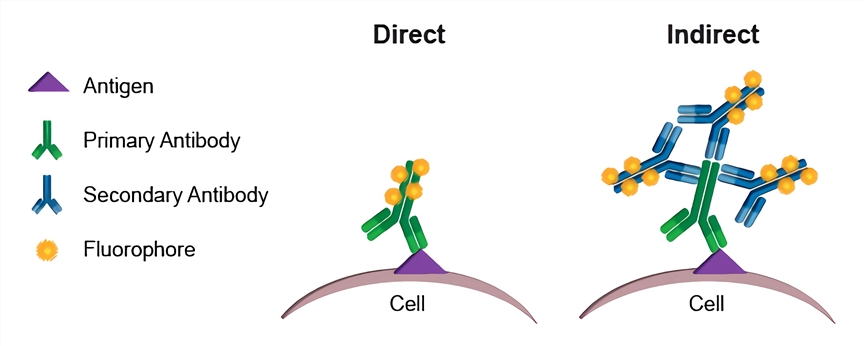
The Intacellular Flow Cytometry Staining Protocol describes the process for intracellular staining of various cell types in vivo-stimulated tissues in vitro-stimulated cultures and whole blood. Direct and indirect staining staining of intracellular antigens permeabilization and cell preparation protocols. If you are unable to immediately read your samples on a cytometer keep them shielded from light and in.
Ad Cidx flow cytometry derived from HEK293 high Purity high batch-to-batch consistency. Flow cytometry FACS staining protocol Cell surface staining 1. Ad Contains Lysing Solution and Fixation Permeabilization Wash Buffers for Flow Cytometry.
Ad Cidx flow cytometry derived from HEK293 high Purity high batch-to-batch consistency. Dilute the fluorochrome-labeled secondary antibody in 3 BSAPBS at the optimal dilution according to the manufacturers instructions and then resuspend the cells in this solution. Ad Compatible with optional autosamplers for walk-away automation and robotic integration.
Run difficult samples at high flow rates with a system that is less sensitive to clogging. Recombinant proteins designed for biological medicine RD. If using the Attune Acoustic.
Add 100 µL of 200 µgmL DNase-free RNaseA and incubate at 37C for 30 minutes. Run difficult samples at high flow rates with a system that is less sensitive to clogging. Wash the cells once with cold PBS at 300-400 x g and re-suspend in.
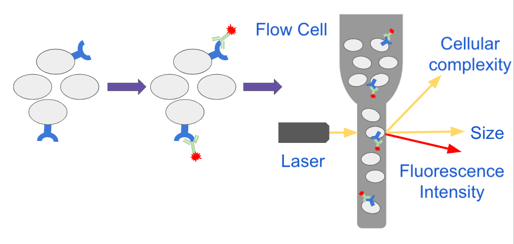
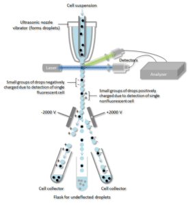
Flow cytometry FACS staining protocol Cell surface staining Harvest wash the cells single cell suspension and adjust cell number to a concentration of 1-5x10⁶ cellsml in ice cold. FACS is an abbreviation for fluorescence-activated cell sorting which is a flow cytometry technique that further adds a degree of functionality. Flow Cytometry Facs Calibur System supplied by Beckman Coulter used in various techniques.
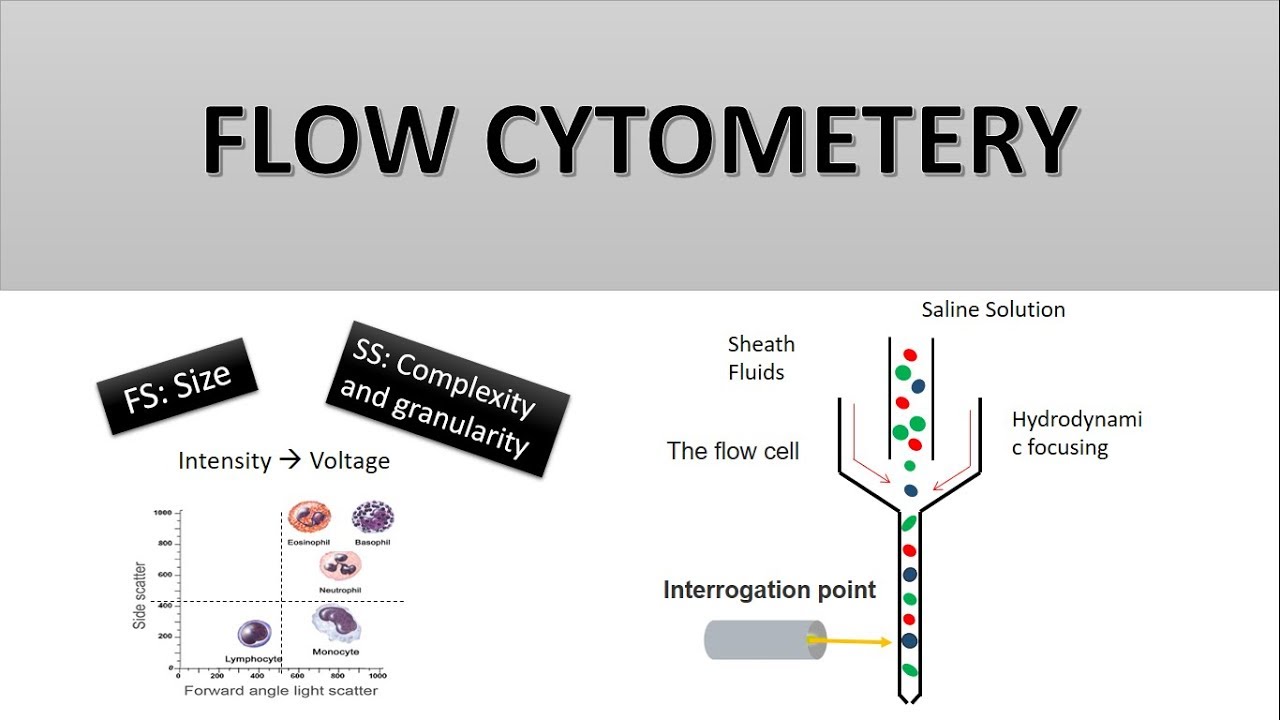
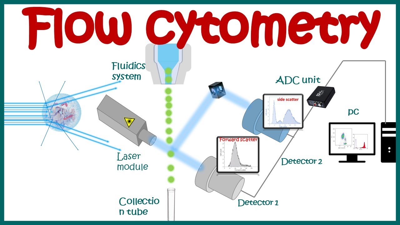
86100 based on 1 PubMed citations. Flow cytometry FCM is a means of measuring certain physical and chemical characteristics of cells or particles as they pass in a fluid stream by a beam of laser light.

Flow Cytometry Facs Protocols Sino Biological
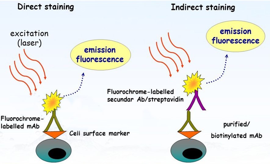
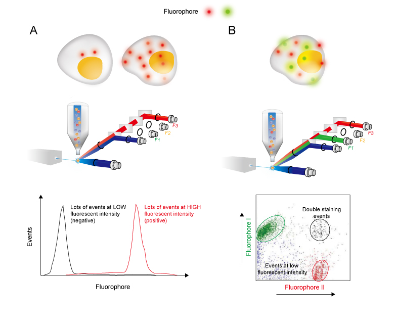
Indirect Staining Flow Cytometry Creative Biolabs

Flow Cytometry Introduction Abcam

Flow Cytometry Protocols

Flow Cytometry Detection Of Surface And Intracellular Antigens In Pancreas From A Single Mouse Embryo Star Protocols
Analyzing Single Cells With Flow Cytometry

In The Protocol Developed By Bernhard Fuchs S Team Bacterial Groups Are Enriched In Three Steps 1 In Situ Hybridization Postdoctoral Researcher Microbiology
Flow Cytometry Guide Creative Diagnostics
How Does Flow Cytometry Work Nanocellect

Flow Cytometry Based Protocols For Human Blood Marrow Immunophenotyping With Minimal Sample Perturbation Star Protocols
The Principle Of Flow Cytometry And Facs 2 Facs Fluorescence Activated Cell Sorting Youtube

Single Cell Rna Expression Analysis Using Flow Cytometry Based On Specific Probe Ligation And Rolling Circle Amplification Acs Sensors
Flow Cytometry Basic Principles What The Use Of Flow Cytometry Cell Sorting By Facs Youtube

Optimized Flow Cytometric Protocol For The Detection Of Functional Subsets Of Low Frequency Antigen Specific Cd4 And Cd8 T Cells Sciencedirect

Flow Cytometry Sample Preparation Proteintech Group
Flow Cytometry Creative Biolabs

Flow Cytometry Creative Biolabs

Flow Cytometry For Dna Analysis Youtube

Diagnostic Potential Of Imaging Flow Cytometry Trends In Biotechnology